
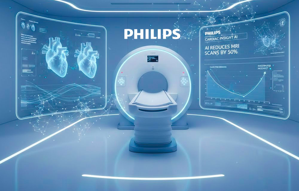
The gold standard for diagnosing heart disease just got twice as fast, infinitely more accessible, and remarkably clearer.
The Netflix-Length Heart Scan Is Finally Here
Imagine lying in an MRI tunnel for what feels like an eternity. Your heart racing. Breath-holding commands echoing through the machine. Forty-five minutes of claustrophobic stillness, all for images that might still come out blurry.
Now imagine getting that same life-saving cardiac scan done in half the time – with image quality so sharp your cardiologist can see details previously hidden in the noise.
This isn’t science fiction. This is what Philips just unveiled at the European Society of Cardiology congress: an AI-powered Cardiac MRI Suite that represents the most significant leap in heart imaging technology since MRI was invented.
And it’s already changing lives.
The Problem: Cardiac MRI Has Been Stuck in the Slow Lane
Cardiac MRI has long been considered the gold standard for evaluating heart disease. It provides unmatched soft-tissue contrast, can measure heart function with precision, and detects problems other imaging modalities miss.
But it has three massive problems:
1. Time. Traditional cardiac MRI scans take 45–60 minutes. Patients must hold their breath repeatedly while remaining motionless. Any movement creates artifacts that blur the images, forcing technicians to repeat sequences.
2. Access. MRI machines are expensive, complex, and require constant maintenance. The 1,500–2,000 liters of liquid helium needed to keep the magnets superconducting evaporate over time, requiring costly refills. A single helium quench – an emergency shutdown where all the helium escapes – can cost hundreds of thousands of dollars and sideline the machine for months.
3. Quality variability. Heart motion, breathing, and patient anxiety all conspire to degrade image quality. Radiologists often have to make diagnoses based on imperfect data.
The result? Massive waiting lists. Limited access in rural areas. Patients avoiding scans due to anxiety. And doctors making life-or-death decisions with less-than-ideal images.
Until now.
The Breakthrough: Deep Learning Meets the Human Heart
Philips’ new AI-powered Cardiac MRI Suite doesn’t just incrementally improve on the old technology. It fundamentally rethinks how cardiac images are created, reconstructed, and enhanced.
At its core is a sophisticated deep-learning system trained on tens of thousands of cardiac cycles from diverse patient populations. The AI has learned to understand the complex, dynamic motion of the human heart – how it contracts, relaxes, and responds to breathing.
Here’s what makes it revolutionary:
Real-Time Motion Prediction and Correction
Instead of treating heart motion as noise to be eliminated, the AI predicts it. As the scan runs, the system anticipates where the heart will be in the next microseconds and adjusts the imaging parameters accordingly. Motion artifacts that would normally ruin scans simply don’t appear.
Intelligent Data Reconstruction
Traditional MRI requires capturing massive amounts of raw data to create clear images. Philips’ AI intelligently fills in gaps, using its deep understanding of cardiac anatomy to reconstruct high-quality images from significantly less data. This is what enables the dramatic speed increase.
Adaptive Enhancement
The system doesn’t just create images – it enhances them in real time. Contrast is optimized. Edges are sharpened. Subtle abnormalities are made visible. All automatically, while the patient is still in the scanner.
The result? Scan times slashed by up to 50%, with image quality radiologists describe as “stunningly sharp.”
A cardiologist at a Munich hospital who participated in early testing put it bluntly: “I zoomed in on a coronary artery segment I normally struggle to visualize. It was like looking through a crystal-clear window instead of frosted glass. I could see details I’ve never seen before.”
The Second Revolution: Nearly Helium-Free MRI Changes Everything
Speed and quality would be impressive enough. But Philips didn’t stop there.
They paired their AI suite with BlueSeal magnet technology – the world’s first and only nearly helium-free MRI system. And this might be even more transformative than the AI itself.
Why Helium Matters More Than You Think
Liquid helium is what keeps MRI magnets at superconducting temperatures near absolute zero. Traditional systems hold 1,500–2,000 liters of the stuff, which slowly evaporates and must be refilled regularly at enormous cost.
Helium is also increasingly scarce. It’s a non-renewable resource primarily extracted as a byproduct of natural gas production. Prices have skyrocketed. Supply disruptions are common. Some hospitals have delayed or cancelled MRI programs simply because they couldn’t secure reliable helium supplies.
And then there are quenches – catastrophic events where the helium explosively boils off. Beyond the massive cost, quenches can render machines unusable for months while repairs are made and the system is refilled.
BlueSeal Changes the Game
Philips’ BlueSeal technology uses just 7 liters of helium – sealed for life. That’s more than 99% less than conventional systems.
The implications cascade through every aspect of cardiac MRI:
- Dramatically lower operating costs (no helium refills, ever)
- No risk of expensive quenches
- Simpler installation (no helium vent pipes through multiple floors)
- Smaller footprint (fits in spaces where traditional MRIs won’t)
- Environmental sustainability (conserving a precious non-renewable resource)
But the most profound impact is on access.
For the first time, world-class cardiac MRI becomes realistic for rural hospitals that lack specialized cryogenic infrastructure. For developing countries where helium supply chains are unreliable or non-existent. For mobile imaging units that bring diagnostics to underserved communities.
A technology once limited to major academic medical centers can now reach the patients who need it most.
Real-World Results Are Already Transforming Care
This isn’t vaporware or a distant promise. Philips’ AI-powered Cardiac MRI Suite is already deployed in early adopter sites across Europe, and the results are remarkable.
Throughput Revolution
A hospital in the Netherlands recently completed 18 full cardiac studies in a single day – a number they previously achieved only once or twice per week. The AI acceleration allowed them to triple their daily capacity without adding staff or extending hours.
Other sites report consistent 30–40% increases in daily cardiac MRI throughput. Waiting lists that stretched months are now measured in days.
Patient Experience Transformed
Shorter scans mean less anxiety. Patients who previously struggled to complete full cardiac protocols – elderly patients, those with back pain, claustrophobic patients – now breeze through examinations.
One radiology technician noted: “We used to have patients leave mid-scan because they couldn’t tolerate it. Now they’re walking out saying ‘That’s it? We’re done already?'”
Diagnostic Confidence Soaring
The enhanced image quality is allowing cardiologists to detect subtle abnormalities they would have missed before. Early signs of cardiomyopathy. Small areas of myocardial scarring. Coronary artery anomalies that previous MRI systems rendered as ambiguous shadows.
A cardiac imaging director in Germany shared: “We’re identifying silent heart conditions in patients who came in for routine screening. Problems that would have gone undetected until they became emergencies. This technology isn’t just making our jobs easier – it’s saving lives.”
Why This Matters More Than Ever
The timing of this breakthrough couldn’t be more critical.
Cardiovascular disease remains the number one killer globally, claiming 18.6 million lives every year. In the United States alone, someone has a heart attack every 40 seconds. Many of these deaths are preventable – if the conditions are caught early enough.
Yet access to advanced cardiac imaging remains profoundly unequal:
- Developing nations have 1/100th the MRI capacity of wealthy countries
- Rural hospitals lack the resources for complex cardiac imaging programs
- Even in developed nations, months-long waiting lists delay diagnosis
- Cost pressures force healthcare systems to ration access to cardiac MRI
Philips’ dual innovation – AI acceleration paired with helium-free operation – directly attacks both the time barrier and the access barrier.
This isn’t an incremental improvement. It’s a democratization of life-saving technology.
For the first time, a community hospital in rural India or Kenya or Peru could realistically operate a world-class cardiac MRI program. Mobile imaging units could bring advanced diagnostics to remote populations. Overwhelmed urban hospitals could double their capacity without building new facilities.
The geographic and economic lottery of cardiac care just got a little less brutal.
The Technology Arms Race: Philips Pulls Ahead
While competitors like Siemens Healthineers and GE Healthcare scramble to develop their own AI-enhanced cardiac imaging solutions, Philips has established a commanding lead.
Their advantage isn’t just technological – it’s strategic.
By integrating AI acceleration with helium-free magnets, they’ve created a complete solution rather than a incremental upgrade. They’re not just making existing systems faster; they’re reimagining what cardiac MRI can be and where it can exist.
The company is rolling out the new AI-powered Cardiac MRI Suite globally throughout 2025 and beyond, with installations already planned across North America, Europe, Asia, and emerging markets.
Early sales figures suggest pent-up demand is enormous. Hospitals that had postponed MRI upgrades due to helium concerns are now pulling the trigger. Facilities that never considered cardiac MRI programs are suddenly reassessing.
The Future: Where Cardiac Imaging Goes Next
If Philips’ breakthrough is any indication, we’re entering a golden age of cardiac diagnostics.
What’s coming next:
AI That Diagnoses, Not Just Images
The next evolution won’t just create better images – it will interpret them. AI systems trained on millions of scans and outcomes will flag abnormalities in real time, suggest differential diagnoses, and even predict future cardiac events based on subtle patterns invisible to human eyes.
Personalized Scanning Protocols
Machine learning will tailor each scan to the individual patient’s anatomy, cardiac rhythm, and clinical history. No more one-size-fits-all protocols that waste time on unnecessary sequences while missing critical details.
Integration with Wearables and Continuous Monitoring
Imagine your smartwatch detecting an irregular heartbeat and automatically triggering a cardiac MRI appointment – with the AI already pre-loaded with your baseline heart data from years of continuous monitoring. Diagnosis and treatment planning could begin before you even know something’s wrong.
Fully Automated Workflows
From patient scheduling to image acquisition to preliminary reporting, AI will streamline the entire cardiac imaging pipeline. Radiologists will shift from spending hours on routine interpretations to focusing on complex cases that require human expertise.
The Human Impact: Stories That Matter
Behind every technological specification is a human story.
There’s the 52-year-old marathon runner whose subtle coronary anomaly was caught during a routine screening – a time bomb discovered and defused before it could detonate.
The 68-year-old woman with severe claustrophobia who finally completed her cardiac MRI after three previous failed attempts – and discovered the early-stage cardiomyopathy that explained her worsening fatigue.
The community hospital in rural Wisconsin that can now offer cardiac MRI for the first time, keeping patients close to home instead of sending them on five-hour drives to the nearest academic medical center.
The cardiologist in Nairobi whose new MRI system doesn’t require impossible-to-obtain helium refills – meaning consistent, reliable cardiac imaging for her patients year after year.
These are the stories that matter. These are the lives that change.
The Bottom Line: A Watershed Moment
Philips hasn’t just improved cardiac MRI. They’ve fundamentally transformed what’s possible in heart diagnostics.
For patients: Faster scans, less anxiety, earlier detection, better outcomes.
For doctors: Images of unprecedented clarity, diagnostic confidence, the ability to see what was previously invisible.
For healthcare systems: Doubled capacity, eliminated helium costs, access for previously underserved populations.
For the world: A powerful new weapon in the fight against humanity’s biggest killer.
The heart just received its biggest upgrade in decades.
And it’s beating faster, stronger, and clearer than ever before.
About This Technology
Manufacturer: Philips Healthcare
Product: AI-Powered Cardiac MRI Suite with BlueSeal Technology
Key Innovation: 50% faster scans using deep-learning motion correction and intelligent image reconstruction
Helium Requirements: 7 liters (sealed for life) vs. 1,500-2,000 liters in conventional systems
Availability: Global rollout ongoing throughout 2025
Impact: Transforming access to life-saving cardiac diagnostics worldwide
Published by VFutureMedia – Exploring Tomorrow’s Technology Today
Follow us for more breakthrough healthcare technology stories that are reshaping medicine and saving lives.
If you found this useful, the best thing you can do is share it with someone who’d actually appreciate it. And if you want more like it, we’re here every week.


Leave a Comment